Superficial brain waves can be easily read from the scalp (see also EEG). These can be altered by various external stimuli. In this examination, a change in brain waves (potentials) is triggered (evoked) by subtle electrical stimuli at nerve endings in the body (somatosensory).
Measurement of sensory evoked potentials examines conduction in the sensory system. This includes the nerves responsible for sensation (feeling, e.g. touch sensation, pressure sensation, etc.) in the legs, arms or face, the sensory nerve root in the spinal area, the nerve fibers in the spinal cord and conduction in the brain to the cerebral cortex, which specifically processes sensation.
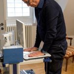
The test is conducted using measuring electrodes which are attached (usually glued) to the scalp at the beginning. The sensitivity stimulus is given as an electrical impulse via a nerve on the leg (rarely on the arm or face). Slight muscle twitches should be visible. The stimulus excites the nerves and is then conducted to the spinal cord. From there, it then travels via circuitry to various centers in the brain and then to the cerebral cortex.
These impulses can be derived and measured via electrodes on the spine or shoulder and on the head. Based on the time sequence and shape of the potentials, conclusions can be drawn about the functioning of the pathways from the nerve endings on the body to the processes in the brain. These potentials may be pathologically altered in some diseases. Abnormal function of sensory nerve pathways can thus be detected and localized, such as in multiple sclerosis or spinal canal narrowing with spinal cord entrapment or other spinal cord diseases.
This method is also well suited for follow-up examinations. Since the sensory evoked potentials are very small and are masked by muscle movements, eye movements, etc., many small stimuli (at least 100 per side) must be applied. It is especially important that the patient is relaxed and does not move. Sources of interference such as hearing aids or cell phones must be switched off.
Often, motor evoked potentials (MEPs) running in the reverse direction in the motor nervous system are also examined as a supplement to SEPs. Both examinations can also be performed by neurologists for continuous monitoring of the nervous system during operations, for example on the spine. These techniques then form part of intraoperative neuromonitoring (IOM).
The examination is harmless and not painful. Occasionally, the electrical impulses are experienced as unpleasant. It takes between 10 and 45 minutes in total.